Polydactyly Of The Foot Repair With Circumferential Racquet-Shaped Incision Technique And Collateral Ligament Reconstruction: Case Report And Literature Reviews
DOI:
https://doi.org/10.14228/jpr.v6i2.282Keywords:
Polydactyly; Circumferential Racquet-Shaped Incision; Collateral Ligament ReconstructionAbstract
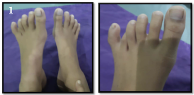
Introduction: Polydactyly of the foot is the most common anomaly in congenital diseases. This abnormality is characterized by the presence of additional digits outside the normal complement of the foot. Polydactyly has reported about 1 in 1,000 births. The Watanabe classification is used in assessing polydactyly in the feet as a fifth ray duplication. Various operating techniques can sometimes be an option. Each method has advantages and disadvantages that can be considered in the selection of surgical in the correction polydactyly.
Patient and Method: we report a case of polydactyly of the foot reconstruction using circumferential racquet-shaped incision, osteotomy and collateral ligament reconstruction that was carried out at Bahteramas Hospital, Kendari. A circumferential racquet-shaped incision design was performed over the sixth MTP joint. Complete excision of the lateral sixth toe was performed, followed by osteotomy of the fifth metatarsal lateral head with a micro-sagittal oscillating saw. The collateral ligament was reconstructed using ligamentoperiosteal flap and fixed using syringe needle to replace K-Wire.
Result : After postoperative control for 3 months the patient was declared good wound healing
Conclusion: Optimal surgical planning, good patient orientation and surgical techniques that are carefully considered are essential for optimal results. Herein, we report a case of polydactyly and racquet shaped incision technique performed to reconstruct the polydactyly.
References
2. Bunnell S. Surgery of the Hand (4th ed). Philadelphia: Lippincott, 1964.
3. Guo, B., Lee, S. K., & Paksima, N. (2013). Polydactyly. Bulletin of the Hospital for Joint Diseases, 71(1).
4. Watanabe H, Fujita S, Oka I. Polydactyly of the foot: an analysis of 265 cases and a morphological classification. PlastReconstr Surg.1992;89:856-877.
5. Galois, L., Mainard, D., & Delagoutte, J. P. (2002). Polydactyly of the foot. Literature review and case presentations. Acta orthopaedica Belgica, 68(4), 376-380.
6. Fortems Y, De Smet L, Fabry G. Extra digit at the dorsum of the foot: hypoplastic central ray metatarsal duplication. J Pediatr Orthop 1996; 5B: 132-3.
7. Rafique A, Arshad A and Abu-Zaid A. Rare presentation of foot postaxial polydactyly. J Foot Ankle Surg 2014; 53: 331–334.
8. Lui TH. Correction of postaxial metatarsal polydactyly of the foot by percutaneous ray amputation and osteotomy. J Foot Ankle Surg 2013; 52: 128–131. 5. Turra S, Gigante C and Bisinella G. Polydactyly of the foot. J Pediatr Orthop B 2007; 16: 216–220.
9. Nakamura K, Ohara K, Ohta E. A new surgical technique for postaxial polydactyly of the foot. Plastic and Reconstructive Surgery. 1996; 97(1): 133–138. DOI:10.1016/S0260-4779(98) 00022-3.
10. Lister, G. (1985). The choice of the procedure following thumb amputation. Clinical Orthopedics and related research, (195), 45-51.
11. Light, T. R. (1992). TREATMENT OF PREAXIAL. Hand clinics, 8(1), 161.
12. Talusan, P. G., Milewski, M. D., & Reach Jr, J. S. (2013). Fifth toe deformities: overlapping and underlapping toe. Foot & ankle specialist, 6(2), 145-149.
13. Baek, G. H. (2015, December). Surgical strategy for polydactyly of the thumb. In BMC proceedings (Vol. 9, No. 3, p. A10). BioMed Central.
Downloads
Published
Issue
Section
License

This work is licensed under a Creative Commons Attribution-NonCommercial-NoDerivatives 4.0 International License.
Authors retain the copyright of the article and grant Jurnal Plastik Rekonstruksi the right of first publication with the work simultaneously licensed under a Creative Commons Attribution License. Articles opting for open access will be immediately available and permanently free for everyone to read, download and share from the time of publication. All open access articles are published under the terms of the Creative Commons Attribution-Non-commercial-NoDerivatives (CC BY-NC-ND) which allows readers to disseminate and reuse the article, as well as share and reuse of the scientific material. It does not permit commercial exploitation or the creation of derivative works without specific permission.